What Is an Orthopaedic Knee Replacement?
An orthopaedic knee replacement is an operation that replaces damaged joint surfaces with artificial components. It may be considered when arthritis or injury causes lasting pain, stiffness, and difficulty with everyday tasks. A short walk can feel long. Stairs may become a daily negotiation.
The procedure is not a new knee in the literal sense. A surgeon removes selected damaged surfaces and fits metal and plastic parts to help the joint move more comfortably. The exact design depends on the person’s knee, health, and activity goals. As orthopaedic surgeon Dr. Daniel J. Berry has explained in patient guidance, the aim is to relieve pain and improve function—not to promise a perfect knee. That distinction matters. Recovery takes time, and results vary.
This guide explains what happens before, during, and after an orthopaedic knee replacement. It also covers the difference between partial and total replacement, common risks, and questions to discuss with a qualified surgeon. A careful assessment may include a physical examination, medical history, and X-rays. The decision should reflect more than a scan. It should consider pain, mobility, overall health, and what matters in daily life. There are trade-offs. Some people regain comfortable movement, while others continue to notice stiffness or limits. Knowing what the operation can—and cannot—do helps patients make a more informed choice with their care team.
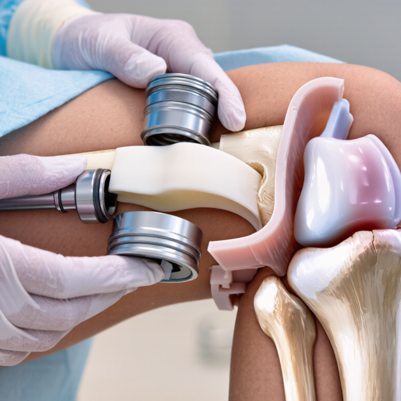
Definition and Purpose of an Orthopaedic Knee Replacement
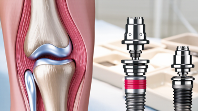
An orthopaedic knee replacement is an operation that replaces damaged parts of the knee joint with artificial components. An orthopaedic surgeon may recommend it when severe arthritis or another condition causes persistent pain and limits everyday movement. During surgery, worn joint surfaces are removed and replaced with components designed to help the knee move more smoothly. The goal is comfort. It is not simply a treatment for every sore knee.
The purpose is to reduce pain and support activities such as walking, standing, and climbing stairs. A replacement may improve function, but it cannot guarantee a pain-free knee or restore every movement. Recovery takes time and usually includes guided exercises to rebuild strength and flexibility. A surgeon considers symptoms, examination findings, imaging, general health, and personal goals before discussing surgery. The decision deserves careful thought.
Tips: Ask what activities are realistic during recovery and how long healing may take. Write down your medicines and health conditions before appointments. Follow your care team’s exercise instructions, but report new swelling, fever, or worsening pain promptly. Recovery can be uneven, and that is worth discussing openly.
Knee Anatomy and Conditions That May Lead to Replacement
The knee is where the thighbone and shinbone meet, with the kneecap guiding movement at the front. Smooth cartilage covers the bone ends and helps them glide. Menisci act as cushions, while ligaments steady the joint. Small structures matter. When cartilage wears away, movement may become painful and stiff.
Osteoarthritis is a common reason people consider knee replacement. It gradually damages cartilage and may cause aching, swelling, or difficulty walking. Inflammatory arthritis can also harm the joint lining and cartilage. A serious fracture or other injury may lead to lasting damage, sometimes years later. The cause is not always simple.
Replacement may be discussed when pain and limited movement disrupt daily activities, and other treatments have not helped enough. A clinician considers symptoms, physical examination, imaging, overall health, and personal goals. An X-ray can show joint changes, but it does not tell the whole story. Pain is not the only measure. The decision should reflect how the knee affects life, not a scan alone. Some people expect one image to provide a clear answer; in practice, the choice can take careful discussion.
Types of Knee Replacement and Their Differences
An orthopaedic knee replacement resurfaces damaged parts of the joint with artificial components. The type depends on where arthritis has damaged cartilage, how stable the knee is, and the person’s overall health. A total knee replacement resurfaces the thighbone and shinbone ends, and may replace the underside of the kneecap. It is commonly considered when damage affects several compartments. A partial knee replacement treats only one damaged compartment, leaving healthier bone, cartilage, and ligaments in place. Recovery can be quicker for some patients, but not everyone’s arthritis is limited enough for this option.
There are differences. A patellofemoral replacement targets damage mainly behind the kneecap; it is less common and suits carefully selected cases. Revision surgery replaces or repairs an earlier implant, often because of wear, loosening, infection, or instability. It can be more complex than a first operation. AHRQ’s HCUP data recorded about 719,000 total knee replacements in U.S. hospitals in 2010, showing how established the procedure is, not which type is best for an individual. The distinction sounds simple. Real knees are not. Imaging, examination, symptoms, and a discussion of likely benefits and risks guide the choice with an orthopaedic clinician.
What Is an Orthopaedic Knee Replacement? Types and Differences
How to read this chart: The knee has three anatomical compartments: medial tibiofemoral, lateral tibiofemoral, and patellofemoral. A total knee replacement resurfaces all three; partial replacements address one or two, leaving unaffected compartments in place. The best option depends on which parts of the knee are damaged and a clinician’s assessment. Revision surgery replaces or repairs a previous implant and can involve a variable number of compartments, so it is not shown as a fixed value.
How the Replacement Procedure Is Performed
An orthopaedic knee replacement resurfaces damaged parts of the knee to reduce pain and improve movement. The procedure is usually performed under spinal or general anaesthesia. The surgical team cleans the skin and makes an incision at the front of the knee. Not a simple swap.
The surgeon moves the kneecap aside and removes worn cartilage and a thin layer of bone from the thighbone and shinbone. Shaped metal components cover these surfaces, with a durable spacer between them to help the joint glide. In a partial replacement, only the damaged compartment is treated. The surgeon checks alignment and knee movement before closing the incision. Exact steps vary with the patient’s anatomy and the surgical plan. The operation ends in the theatre; recovery does not.
Tips: Ask your care team which type of replacement is planned, how your medicines should be managed, and when to begin exercises. Follow the individual instructions for wound care and walking aids. Small details matter, but progress can feel uneven. A stiff morning or a slower day does not automatically mean something is wrong; contact your clinician about concerning symptoms or unexpected changes.
What Is an Orthopaedic Knee Replacement? - How the Replacement Procedure Is Performed
| Procedure stage |
What typically happens |
Purpose or key detail |
| 1. Preparation and anaesthesia |
The surgical team reviews the patient’s health, confirms the operative knee, and prepares the skin. Anaesthesia may be general or regional, such as a spinal anaesthetic; pain-relief measures may also be used. |
The anaesthetic plan is selected for the individual in consultation with the anaesthesia team. |
| 2. Surgical access |
The surgeon makes an incision at the front of the knee and moves the kneecap aside as needed to reach the joint surfaces. |
The incision and approach vary according to the patient’s anatomy and the surgeon’s technique. |
| 3. Damaged surface preparation |
Damaged cartilage and a small amount of underlying bone are removed from the ends of the thighbone (femur) and shinbone (tibia). The back of the kneecap may also be resurfaced. |
The preparation creates surfaces for the replacement components; whether the kneecap is resurfaced depends on the case. |
| 4. Component positioning |
A metal component is fitted to the end of the femur, and a component is fitted to the top of the tibia. A smooth plastic bearing is placed between them; a plastic surface may also be used on the kneecap if resurfaced. |
Components may be fixed with bone cement or, in selected cases, by a method that allows bone to grow onto the component. |
| 5. Alignment and movement checks |
The surgeon checks the fit, stability, alignment, and movement of the knee through a range of motion, making adjustments if needed. |
The aim is a stable joint with appropriate movement and balanced surrounding soft tissues. |
| 6. Closure and dressing |
The surgical area is washed, the incision is closed with sutures, staples, or another closure method, and a dressing is applied. |
The closure method and dressing depend on the surgical team’s practice and the patient’s needs. |
| 7. Early recovery and rehabilitation |
After surgery, the care team monitors recovery and provides pain management. Many patients begin standing or walking with assistance and start exercises soon after the procedure. |
Rehabilitation and recovery timelines vary; follow-up and exercise plans are tailored by the treating team. |
General information only. The exact procedure, risks, and recovery plan vary by patient; discuss individual care with an orthopaedic surgeon.
Recovery, Rehabilitation, and Long-Term Care
After an orthopaedic knee replacement, recovery usually starts in hospital or a supervised care setting. Your team will guide safe transfers, walking, and pain control. Follow their instructions for wound care and medication; plans differ between patients. Keep the incision clean and watch for increasing redness, drainage, fever, or worsening pain. Contact your care team if these occur.
Rehabilitation often begins soon after surgery, with gentle bending, straightening, and short walks. A physical therapist can adjust exercises to your strength and progress. Small steps matter. Use a walker or cane as directed, even when the kitchen seems close. Swelling and stiffness can fluctuate, and progress is rarely a straight line. Rest between sessions, but avoid stopping movement without discussing it with your clinician. Recovery can feel uneven.
At home, clear loose rugs and keep frequently used items within easy reach. As strength improves, your therapist may add balance and leg-strengthening exercises. Do not rush driving, work, or higher-impact activities; ask your surgeon when they are appropriate. Long-term care includes staying active with suitable low-impact exercise, maintaining a healthy weight, and attending recommended follow-up visits. Some days may still feel awkward. That is worth mentioning at checkups, especially if pain or reduced movement persists.